

Developing a New Technology to “Diagnose” Cells by the Color of Light —Successful Visualization of Molecular Condensates in Living Cells and Detection of Cancer‑Related Abnormal States—
Sep 16, 2026
A cross‑disciplinary research group led by associate professor Masaharu Hazawa of the Institute for Frontier Science Initiative, professor Takahiro Soeta of the Faculty of Chemistry, Institute of Science and Engineering, and professor Richard Wong of the Nano Life Science Institute (WPI‑NanoLSI), Kanazawa University, in collaboration with Nagoya University, National Cheng Kung University (Taiwan), and the University of Southern California (USA), has successfully developed “PyLUMI”, a fluorescent probe capable of visualizing the microenvironment inside “molecular condensates” in living cells with unprecedented brightness.
Inside cells, biomolecules such as proteins form structures called biomolecular condensates(*1), without membrane boundaries. These condensates play essential roles in biological processes such as gene expression and cell division. However, methods for examining the internal environment of condensates without destroying living cells have been limited. By innovating the molecular structure of the probe, the research group significantly improved its fluorescence efficiency in aqueous environments, leading to the development of the new probe “PyLUMI”.
Using PyLUMI, the team demonstrated that clear images can be obtained with only small amounts of probe and short exposure times, minimizing cellular damage. This enabled high‑precision imaging of the centrosome(*2), the initiation site of cell division. As a result, they revealed for the first time worldwide that the state of the centrosome changes as the cell cycle progresses. Furthermore, in cells mimicking cancer‑related centrosome amplification, PyLUMI successfully detected abnormal states that cannot be identified by size alone.
These findings are expected to contribute to future diagnostic markers and drug‑development support technologies for diseases involving abnormalities in molecular condensates, including cancer.
This research was published online in the international journal “Advanced Science“ on July 16, 2026 (11:00 PM JST).
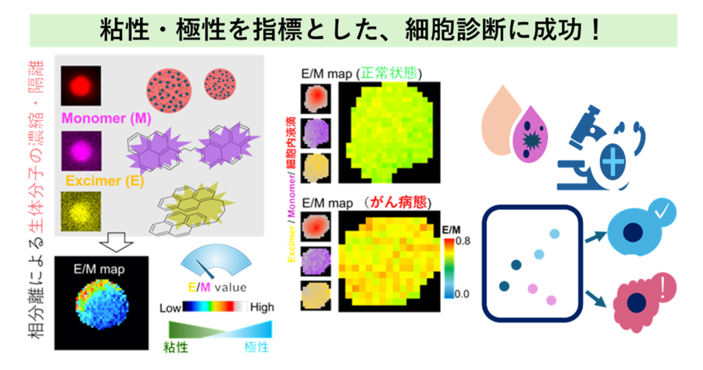
Figure: Visualization of Intracellular Environments Using PyLUMI and Its Application to Cancer Cell Diagnosis
By color‑coding the ratio of excimer emission (E) to monomer emission (M) of two pyrene molecules (E/M ratio) for each pixel, PyLUMI visualizes the viscosity and polarity inside biomolecular condensates. Compared with normal cells (top), cancer‑mimicking cells (bottom) show an overall increase in the E/M ratio, enabling detection of abnormal cellular states that cannot be identified by size alone.
(Modified from K. Matumoto et al., under CC BY 4.0)
【Glossary】
*1 Biomolecular Condensates
Structures formed when biomolecules such as proteins and nucleic acids locally concentrate through liquid–liquid phase separation (LLPS) without membrane boundaries. Examples include nucleoli, stress granules, and centrosomes.
*2 Centrosome
A cellular structure that regulates microtubule formation and serves as the initiation site of spindle formation during cell division. In cancer cells, centrosomes often increase abnormally (centrosome amplification), which is associated with chromosomal instability and cancer malignancy.
Click here to see the press release.【Japanese only】
Journal : Advanced Science
DOI:10.1002/advs.76419
Researcher Information : Masaharu Hazawa
Takahiro Soeta
Richard Wong